Committee I

Committee IV

Renal Tumors

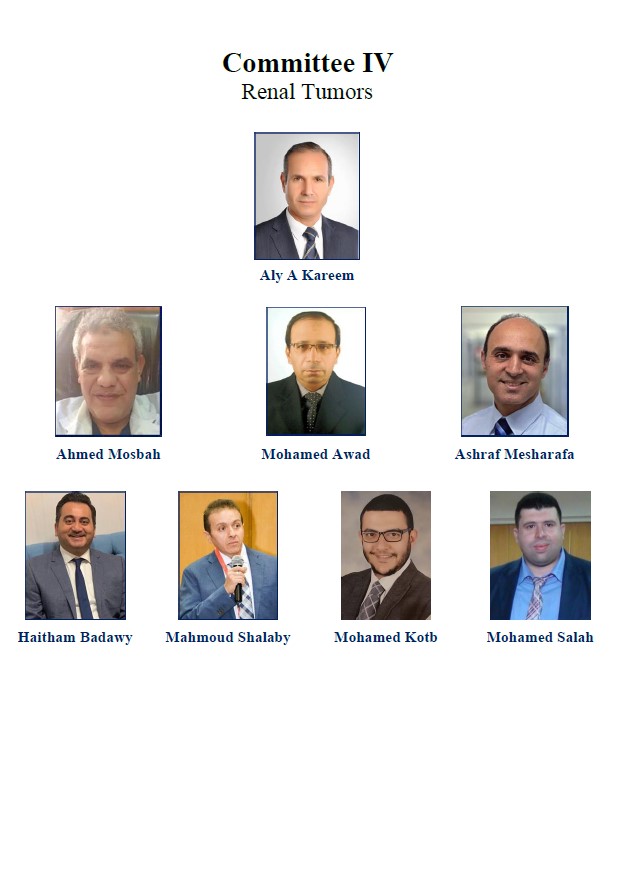
Prof. Aly M. Abdel-Kareem, Professor of Urology.Alexandria University

Prof. Ahmed Mosbah, Professor of Urology.Mansoura University

Prof. Mohamed Awad, Professor of Urology.Mansoura University

Prof. Ashrah Mesharafa, Professor of Urology. Cairo University

Prof. Haytham Badaway, Professor of Urology. Alexandria University

Dr. Mahmoud Shalaby, Assistant Professor of Urology. Assuit University

Dr. Mohamed Kotb, Lecturer of Urology, Ain Shams University

Dr. Mohamed S Elderey, Lecturer of Urology, Zagazig University

Contents
IV.1 List of Abbreviations
  • CT - Computed Tomography
  • CN - Cytoreductive nephrectomy
  • HNPCC - Hereditary Non-Polyposis Colorectal Carcinoma
  • KSS - Kidney Sparing Surgery
  • LNs - Lymph Nodes
  • PN - Partial Nephrectomy
  • PS - Performance Status
  • RN - Radical Nephrectomy
  • RCC - Renal Cell Carcinoma
  • RMB - Renal Mass Biopsy
  • SCC - Squamous cell carcinoma
  • TS - Tuberous Sclerosis
  • US - Ultrasound
  • UTUCs - Upper Urothelial Tract Tumors

IV.2 Introduction

We have been trying to merge updated international guidelines of renal tumors, taking in account the social and financial limitation. It is a step for more development of pure Egyptian guidelines, by also putting solutions for many problems facing us like data registry, multicenter cooperation, and financial limitation.

IV.2.1 Methodology
Data resources included the following:
  • 1. 4 guidelines with their recent updates: National Institute for Health and Care Excellence, European Urological Association, American Urological Association and National Comprehensive Cancer Network.
  • 2. Associated Egyptian publications

The Strength Rating of grading depends on 2 parameters:
Strength Rating of clinical practice recommendation, graded as strong or weak. This is the consensus of the Egyptian guidelines committee for renal tumors, relying on international guidelines recommendations, taking in accounts the socio-economic factors and the Egyptian environment.

IV.2.2 Results
These guidelines represent the best clinical evidence-based medicine available. These guidelines are not solely based upon the level of evidence of the international publications but also include expertise of the treating doctor and expectations of every patient.

IV.2.3 Conclusions.
These guidelines provide practical evidence-based guidance on the clinical aspect of Renal tumors. The main target is entirely focused on assessment and treatment, reflecting the recommended clinical practice. This can provide the basis for thinking through patient’s management and also for planning and designing clinical services.

IV.3 Renal Cell Carcinoma (RCC):
IV.3.1 Epidemiology of Renal Cell Carcinoma (RCC):
Renal cell carcinoma represents around 3% of all cancers worldwide, with the highest incidence occurring in Western countries. Generally, during the last two decades, there has been an annual increase of about 2% in the RCC incidence worldwide. (1)

Based on the results published from the National Cancer Registry Program (NCRP) in 2014, the crude rate for renal cancer was 1.8 while the age-standardized incidence rate was 2.7 for a relative percentage of 1.53% of cancers at all sites in males; while in females it was 0.97%. (2)

20% of Small renal mass less than 4 cm are benign tumors. Oncocytoma is a benign tumor that originates from intercalated collecting duct cells like chromophobe RCC. It is characterized by Y chromosomal anomalies and associated incidence of RCC. It is a well-circumscribed tumor with central stellate scar. On microscopic examination it shows eosinophilic cells with negative Hale stain. As it is difficult to diagnose it clinically, it is diagnosed pathologically post-surgery. (3)

AML (angiomyolipoma) is a benign mesenchyme tumor. It is either sporadic or familial like tuberous sclerosis (TS). It is diagnosed clinically by CT by the presence of fat except in poor fat AML. Venous thrombosis is a rare complication especially in the epithelioid pattern. Familial AML and large size AML have a higher risk for bleeding. (3)

Table IV:1 Recommendations of Epidemiology of RCC

Recommendations

Strength Rating

1.Treat AML either with NSS or angioembolization:
  • a) Tumor > 4cm
  • b) Female in childbearing period.
  • c) Patient away from medical service.
Weak
2. Offer systemic therapy with mTOR Inhibitors for patient not fit for surgery or embolization. Weak
IV.3.2 Etiology of RCC
Factors that have been identified as risk factors in the etiology of RCC include smoking, obesity, and hypertension. Increasing physical activity, eliminating cigarette smoking and in obese patients reducing weight are therefore potential preventative measures to decrease the risk of RCC.(1)

Table IV:2 TNM staging

T - Primary tumor(3)

TX Primary tumour cannot be assessed

T0 No evidence of primary tumour

T1 Tumour ≤ 7 cm or less in greatest dimension, limited to the kidney

T1a Tumour ≤ 4 cm or less

T1b Tumour > 4 cm but ≤ 7 cm

T2 Tumour > 7 cm in greatest dimension limited to the kidney

T2a Tumour > 7 cm but ≤ 10 cm

T2b Tumour > 10 cm, limited to the kidney

T3 Tumour extends into major veins or perinephric tissues but not into the ipsilateral adreal gland and not beyond Gerota fascia

T3a Tumour grossly extends into the renal vein or its segmental (muscle- containing) branches, or tumour invades perirenal and/or renal sinus fat (peripelvic fat), but not beyond Gerota fascia

T3b Tumour grossly extends into the vena cava below diaphragm

T3c Tumour grossly extends into vena cava above the diaphragm or invades the wall of the vena cava

T4 Tumour invades beyond Gerota fascia (including congtiguous extension into the ipsilateral adrenal gland)

N - Regional lymph nodes

NX Regional lymph nodes cannot be assessed

N0 No regional lymph node metastasis

N1 Metastasis in a single lymph node 2 cm or less in the greatest dimension

M - Distant metastasis

M0 No distant metastasis

M1 Distant metastasis

pTNM stage grouping

Stage I T1 N0 M0
Stage II T2 N0 M0
Stage III T3 N0 M0
T1, T2, or T3 N1 M0
Stage IV T4 Any N M0
Any T Any N M1
IV.3.3 Evaluation of Renal masses
IV.3.3.1 Diagnosis and Staging:
IV.3.3.1.1 Symptoms:

Renal masses remain asymptomatic until the late stages. More than 60% of RCCs are detected incidentally with abdominal ultrasound (US) or computed tomography (CT) performed for other reasons). The classic triad of flank pain, visible haematuria, and palpable abdominal mass is rare today, and correlates with advanced disease and subtypes associated with poor prognosis. Paraneoplastic syndromes are found in approximately 30% of patients with symptomatic RCCs. A few patients present with symptoms caused by mRCC, such as bone pain, deterioration of performance status (PS), or persistent cough.(6)

IV.3.3.1.2 Imaging:

For a solid or complex cystic renal mass, high quality, multiphase, cross-sectional abdominal imaging should be performed to optimally characterize and clinically stage the renal mass. Characterization of the renal mass should include assessment of tumor complexity, degree of contrast enhancement (where applicable), and presence or absence of fat .(7) With solid RMs, the most important criterion for differentiating malignant lesions is the presence of contrast enhancement or restriction. CT and MRI cannot reliably distinguish oncocytoma and fat-free angiomyolipoma from malignant renal neoplasms(6).

In patients with RCC, chest CT is the most accurate investigation to diagnose lung metastases or enlarged mediastinal lymph nodes (LNs) Since most bone and brain metastases are symptomatic at diagnosis, bone or brain imaging is performed on indication (6).

In the case of a renal cystic mass, the Bosniak classification distinguishes five categories based on CT presentation, can predict the risk of malignancy, and provides guidance for management (6).

IV.3.3.1.3 Laboratory investigations:

In patients with suspected renal malignancy, physicians should obtain comprehensive metabolic panel, complete blood count, and urine analysis. For patients with a solid or complex cystic renal mass, physicians should assign CKD stage based on GFR and degree of proteinuria.(7).

IV.3.3.1.4 Renal mass biopsy (RMB):

Renal mass biopsy should be considered when a mass is suspected to be hematologic, metastatic, inflammatory, or infectious.(7). Percutaneous renal mass biopsies are increasingly used for histological diagnosis to avoid unnecessary surgery in the event of a benign lesion, to select patients for surveillance, and to obtain histology before ablative treatment. Tumor biopsies are also used in mRCC for the selection of medical and surgical treatment (6).

When considering the utility of RMB, patients should be counseled regarding rationale, positive and negative predictive values, potential risks and non-diagnostic rates of RMB. In the setting of a solid renal mass, RMB is not required for: 1) young or healthy patients who are unwilling to accept the uncertainties associated with RMB; or 2) older or frail patients who will be managed conservatively independent of RMB findings (7). Core biopsies are preferable to fine needle aspiration for solid RMs and are not recommended for cystic RMs due to their low diagnostic yield, unless areas with a solid pattern are present (6).

A core biopsy should be performed with 18G needle and a coaxial technique to minimize the risk of seeding. At least two quality cores (non-fragmented, >10 mm in length) should be obtained, and necrotic areas should be avoided to maximize diagnostic accuracy. Peripheral biopsies are preferable for larger masses, to avoid central necrosis (6).

In experienced centers, percutaneous core biopsies have low morbidity, a high diagnostic yield, and accuracy for the diagnosis of malignancy and RCC type. However, they are non-diagnostic in 2.5–22% of cases. If a biopsy is non-diagnostic, a second biopsy or surgical exploration should be considered (6).

IV.3.3.1.5 Counseling:

In patients with a solid or Bosniak 3/4 complex cystic renal mass, a urologist should lead the counseling process and should consider all management strategies. A multidisciplinary team should be included when necessary. Physicians should provide counseling that includes current perspectives about tumor biology and a patient-specific risk assessment inclusive of sex, tumor size/complexity, histology (when obtained), and imaging characteristics.(7).

During counseling of patients with a solid or Bosniak 3/4 complex cystic renal mass, physicians must review the most common and serious urologic and non-urologic morbidities of each treatment pathway and the importance of patient age, comorbidities/frailty, and life expectancy. (7).

Physicians should review the importance of renal functional recovery related to renal mass management, including the risk of progressive CKD, potential short- or long-term need for renal replacement therapy, and long-term overall survival considerations. Physicians should consider referral to nephrology in patients with a high risk of CKD progression. Such patients may include those with eGFR less than 45 ml/min/1.73m2, confirmed proteinuria, diabetics with preexisting CKD, or whenever eGFR is expected to be less than 30 ml/min/1.73m2 after intervention. (7).

Physicians should recommend genetic counseling for all patients ≤ 46 years of age with renal malignancy and consider genetic counseling for patients with multifocal or bilateral renal masses, or if personal or family history suggests a familial renal neoplastic syndrome (7).

Table IV:3 Recommendations of Diagnostic Evaluation of Renal Tumours

Recommendations

Strength Rating

1. Multiphasic contrast enhanced computed tomography of abdomen and chest is mandatory for diagnosis and staging Strong
2. Bone scan and PET CT is routinely used for staging Weak
3. Performing renal biopsy is indicated before ablative or systemic therapy with coaxial core biopsy technique Strong
4. In cystic renal mass biopsy, should be avoided Strong
5. Bosniak type 4 should be considered a malignant RCC Strong
IV.3.4 Treatment of localised RCC and local treatment of mRCC
Surgical treatment Surgery is the only curative treatment for localised RCC. Based on oncological and functional outcomes, localised T1a-b tumours are best managed by partial nephrectomy (PN) rather than by radical nephrectomy (RN), irrespective of the surgical approach. In clinically localised RCCs of ≤4 cm, compared with RN, PN was associated with equal or better survival, whereas serious adverse event rates, CSS, and time to recurrence were similar for both groups (8)

The decision process is necessarily influenced by surgeon’s factors (own surgical expertise and prior outcomes, as well as comfort with various surgical procedures. Also complexity according to RENAL score is important factor.(9) RN is preferred when all of the following criteria are met: (a) there is high tumor complexity and PN would be challenging, even in experienced hands; (b) there is no preexisting CKD or proteinuria; and (c) the contralateral kidney is normal and the new baseline estimated glomerular filtration rate will likely be greater than 45 mL/min/1.73 m2 .(7)

Partial nephrectomy should be considered for patients with solid or Bosniak 3/4 complex cystic renal masses who are young, and have multifocal masses or comorbidities that are likely to impact renal function in the future, such as moderate to severe hypertension, diabetes mellitus, recurrent urolithiasis or morbid obesity.(7)

Negative surgical margins should be a priority. The extent of normal parenchyma removed should be determined by surgeon discretion considering the clinical situation, tumor characteristics including growth pattern and interface with normal tissue. Tumor enucleation should be considered in patients with familial RCC, multifocal disease or severe CKD to optimize parenchymal mass preservation.(7)

Table IV:4 Recommendations of Treatment of RCC

Recommendations

Strength Rating

1. Partial nephrectomy is indicated in T1 tumors (open/laparoscopy) Strong
2. Laparoscopic / open radical nephrectomy for T2 Tumors and localized tumors difficult to be treated with partial nephrectomy. Strong
3. Minimal invasive approach should be avoided if it will comprise the functional or the oncological outcome. Strong
4. Ipsilateral adrenalectomy is indicated if there is clinical invasion for adrenal gland Strong
5. Offer active surveillance or ablative therapy for comorbid patients with small renal mass Weak
6. Offer embolization as palliative treatment (inoperable cases) in cases of hematuria or flank pain Weak
7. Lymphadenectomy can be done to radiologically positive lymph nodes for staging and therapeutics benefits Weak
8. In non-metastatic locally advanced RCC with venous thrombus. Removal of the tumor with the thrombus is necessary. Strong
9. Don't offer adjuvant therapy for non-metastatic RCC Strong
IV.3.4.1 Cytoreductive nephrectomy (CN):
It is curative only if all tumor deposits can be resected. In patients with multiple metastases it is only palliative and systemic treatment are necessary. IMDC (the metastatic renal cancer database consortium) or MSKCC(Memorial Saloan Kettering Cancer Center) for classifying metastatic patients.(1)

Table IV:5 Recommendation on Treatment of mRCC

Recommendations

Strength Rating

1. CN is not indicated for poor risk MSKCC metastatic patients Strong
2. Start systemic therapy 1st for intermediate risk MSKCC patients whom have no complaint from 1ry tumor. Weak
3. Perform immediate CN in good performance patients whom do not require systemic therapy or in patients with resectable oligometastasis. Weak
4. Offer local treatment for symptoms palliation like metastatectomy or streostatic radiotherapy for brain and bone metastasis Weak
IV.3.4.2 Systemic therapy for advanced disease:
IV.3.4.2.1 Clear cell RCC

IV.3.4.2.1.1 Recommendations for first line treatment:

1- Limited disease burden in asymptomatic patient:
Active surveillance
2- IMDC favorable risk:
Pembrolizumab/axitinib
Sunitinib
Pazopanib
3- IMDC intermediate/poor risk:
A- Immune checkpoint inhibitors available:
Pembrolizumab/axitinib
Ipilimumab/Nivolumab
B- Immune checkpoint inhibitors unavailable:
Sunitinib or Pazopanib

IV.3.4.2.1.2 Recommendations for second line treatment:

1- Prior TKI:
Nivolumab:
Immunotherapy unavailable: Axitinib
2- Prior immune checkpoint inhibitor
Sunitinib or
Pazopanib

IV.3.4.2.1.3 Non-clear cell RCC:

Sunitinib (preferred)
Everolimus

Surveillance protocol post-RN:

The aim of surveillance mainly is to detect recurrences and many classifications system is used to stratify risk of recurrence like university of California system. Also surveillance is used for monitor post-operative complications and functional outcomes. (1)

Table IV:6 Surveillance protocol post-RN

Risk Profile

Surveillance

6 mo 1y 2y 3y 3y
Low US CT US CT CT once every 2 years. Counsel about recurrence risk of 10%
Intermediate/High CT CT CT CT CT Once every 2 years
IV.4 Upper urothelial tract tumors (UTUCs)
IV.4.1 Epidemiology:
UTUCs are not common and represent about 5-10% of urothelial carcinoma. Pelvicalyceal tumors are more common than ureteral tumor, 60% of UTUCs are invasive. Concurrent bladder cancer occurs in 17% of cases while up to 47% of cases show bladder recurrence. Familial/hereditary UTUCs are linked to hereditary non-polyposis colorectal carcinoma (HNPCC).(10)

IV.4.2 Risk factors:
Exposure to tobacco or aromatic amines are risk factors to develop UTUCs. Aristolochic acid contained in Aristolochia fangchi and Aristolochia clematis is linked to P53 mutation which increased risk of UTUCs as in Balkan nephropathy. (10)

IV.4.3 Histologic Variants:
Urothelial carcinoma is the most common variant. Squamous cell carcinoma accounts for 10% only. SCC usually associated with chronic inflammation and infection. (10)

Table IV:7 TNM staging

T - Primary tumor (11)

TX Primary tumour cannot be assessed

T0 No evidence of primary tumour

Ta Non-invasive papillary carcinoma

Tis Carcinoma in situ

T1 Tumour invades subepithelial connective tissue

T2 Tumour invades muscle

T3 (Renal pelvis) Tumour invades beyond muscularis into peripelvic fat or renal parenchyma (Ureter)

Tumour invades beyond muscularis into periureteric fat

T4 Tumour invades adjacent organs or through the kidney into perinephric fat

N - Regional lymph nodes

NX Regional lymph nodes cannot be assessed

N0 No regional lymph node metastasis

N1 Metastasis in a single lymph node 2 cm or less in the greatest dimension

N2 Metastasis in a single lymph node more than 2 cm but not more than 5 cm in the greatest dimension or multiple lymph nodes, none more than 5 cm in greatest dimension

N3 Metastasis in a lymph node more than 5 cm in greatest dimension

M - Distant metastasis

M0 No distant metastasis

M1 Distant metastasis

Use the WHO 1973 and 2004 grading systems for the histological classification of UTUC

Table IV:8 Recommendations for Diagnosis of UTUCs

Recommendations (10)

Strength Rating

1. CT urography and urinary cytology is essential for diagnosis Strong
2. Cystoscopy is indicated to diagnose associated bladder tumors. Strong
3. Diagnostic ureterscopy, biopsy and retrograde ureteropyelogram is indicated when additional information will impact treatment strategy. Weak
Table IV:9 Risk stratifications of UTUCs

Low risk (all factors should present)

High risk (any factor present) (10)

Unifocal disease
Tumour size < 2 cm
Low-grade cytology
Low-grade URS biopsy
No invasive aspect on CTU-urography

Hydronephrosis
Tumour size > 1 cm
High-grade cytology
High-grade URS biopsy
Multifocal disease
Previous radical cystectomy for bladder cancer

Table IV:10 Recommendations for Disease management of localized tumors

Recommendations (10)

Strength Rating

1. Kidney sparing surgery (KSS) should be offered for low-risk disease Strong
2. KSS is offered for distal ureteral tumor or in patient with solitary kidney or poor renal function, but thus should not affect oncological outcomes. Weak
3. Radical nephroureterectomy with removal of bladder cuff is indicated in high-risk patient. Strong
4. Lymphadenectomy is performed in high-risk patients Weak
5. Offer intravesical chemotherapy to lower bladder recurrence. Strong
The instillations of BCG post KSS still have not been confirmed to be beneficial. (10)

Laparoscopic radical nephroureterctomy should avoided in cT3/4 or N positive tumors (10)

Metastatic disease:

Radical nephroureterctomy has no role in metastatic disease. It may be offered as palliative treatment. Platinum based chemotherapy should be offered but still data is insufficient. Radiotherapy has no role. (10)

IV.4.3.1 After Radical Nephroureterectomy:
  • low risk
  • Perform cystoscopy at 3month and 12 month then yearly for 5 years.
  • high risk
  • Cystoscopy and cytology every 3months for 2years then every 6 months till 5 years then yearly CT urography and CT chest every 6 months for 2 years then every year.

IV.4.3.2 After kidney Sparing Surgery
  • Low risk
  • Perform cystoscopy and CT urogram at 3month and 6 month then yearly for 5 years.
  • Perform uretescopy at 3 months
  • High risk
  • Cystoscopy and cytology and CT urograpgy and CT chest at 3and 6 month then yearly
  • Ureterscopy and cytology in situ at 3month and 6 month.

IV.4.4 References:
1. Ljungberg B, Albiges L, Abu-Ghanem Y, Bensalah K, Dabestani S, Fernández-Pello S, et al. European association of urology guidelines on renal cell carcinoma: the 2019 update. European urology. 2019; 75(5):799-810.
2. Ibrahim AS, Khaled HM, Mikhail NN, Baraka H, Kamel H. Cancer incidence in Egypt: results of the national population-based cancer registry program. Journal of cancer epidemiology. 2014; 2014.
3. Moch H, Cubilla AL, Humphrey PA, Reuter VE, Ulbright TM. The 2016 WHO classification of tumours of the urinary system and male genital organs—part A: renal, penile, and testicular tumours. European urology. 2016; 70(1):93-105.
4. Brugarolas J. Molecular genetics of clear-cell renal cell carcinoma. Journal of clinical oncology. 2014; 32(18):1968.
5. Nagi FM, Omar A-AM, Mostafa MG, Mohammed EA, Abd-Elwahed Hussein MR. The expression pattern of Von Hippel-Lindau tumor suppressor protein, MET proto-oncogene, and TFE3 transcription factor oncoprotein in renal cell carcinoma in Upper Egypt. Ultrastructural pathology. 2011; 35(2):79-86.
6. Ljungberg B, Albiges L, Abu-Ghanem Y, Bensalah K, Dabestani S, Fernandez-Pello S, et al. European Association of Urology Guidelines on Renal Cell Carcinoma: The 2019 Update. Eur Urol. 2019;75(5):799-810.
7. Campbell S, Uzzo RG, Allaf ME, Bass EB, Cadeddu JA, Chang A, et al. Renal Mass and Localized Renal Cancer: AUA Guideline. J Urol. 2017; 198 (3):520-9.
8. MacLennan S, Imamura M, Lapitan MC, Omar MI, Lam TB, Hilvano-Cabungcal AM, et al. Systematic review of oncological outcomes following surgical management of localised renal cancer. European urology. 2012; 61(5):972-93.
9. Kim JK, Lee H, Oh JJ, Lee S, Hong SK, Lee SE, et al. Comparison of robotic and open partial nephrectomy for highly complex renal tumors (RENAL nephrometry score≥ 10). PloS one. 2019;14 (1):e0210413.
10. Rouprêt M, Babjuk M, Burger M, Capoun O, Cohen D, Compérat EM, et al. European association of urology guidelines on upper urinary tract urothelial carcinoma: 2020 update. European urology. 2020.
11. Sobin L, Wittekind C. Classification of Malignant Tumours TNM. International Union against Cancer UICC. 2009; 6:131-41.

IV.5 WILMS’ TUMOUR “Nephroblastoma”
IV.5.1 Epidemiology
It is the 5th most common tumor, the 2nd common abdominal tumor and the 1st common renal tumor in children. It represents 5% of pediatric tumors. Bilateral wilms occur in 5-7% of patients. More than 80% occur before 5yrs of age with highest incidence among blacks & lowest among Asians

IV.5.2 Pathology:
IV.5.2.1 Microscopic:
1) “Classic” Wilms’

It is composed of blastemal cells, epithelial cells, & stromal cells, If mostly blastemal (highly aggressive tumours) will responds to chemotherapy. However, if mostly epithelial will be less aggressive but not will respond to chemotherapy. Kids less than 1yr of age diagnosed with Wilms’ tumor with Nephrogenic Rests are associated with increased risk of developing contralateral disease

2) Anaplastic Wilms’

It is characterized by 3 histologic abnormalities, nuclear enlargement (3-4x adjacent cells), hyperchromasia of enlarged nuclei and abnormal mitotic figures. Presence of diffuse anaplasia associated with resistance to chemotherapy with worst prognosis.

IV.5.3 Staging
  • COG Staging system used for Wilms’ tumour: (Children’s Oncology Group)
  • Based on surgical & histopathologic findings

Table IV:11 Staging system for Wilm’s Tumour

Stage

Capsule

Surgical margin

Tumour

Hilar structure

I

Limited to kidney

intact renal capsule

-ve completely excised

no rupture

II

not limited to kidney

beyond renal capsule

-ve completely excised

Focal spillage

+/- extrarenal vessel thrombus

III

Residual tumour confined to abdomen

+ve Incomplete excision

Previous Biopsy. Diffuse spillage /rupture peritoneal implant

+ve LNs

IV

Hematogenous metastasis

V

BILATERAL

IV.5.4 Diagnosis:
Abdominal mass, gross hematuria is common presentation. The child seems well unlike neuroblastoma. Ultrasound determines solid and cystic renal mass. Doppler ultrasound can detect renal vein thrombosis. Cross sectional imaging on abdomen and chest should be used for Wilms tumor staging.

Table IV:12 Recommendations for Diagnosis of Wilms Tunour

Recommendation

Strength Rating

1. Cross sectional imaging on abdomen and chest for Wilms tumor staging Weak
2. Bone scan and skeletal survey for bone metastasis Weak
3. Histopathological examination should be combined with chromosomal evaluation to set the management plan Weak
IV.5.5 Treatment
IV.5.5.1 Principles of Surgical Management:
  • Radical Nephrectomy with sampling of suspicious LNs
    • Trans peritoneal approach assess entire abdomen liver, LN for Mets.
    • No role for exploration of contralateral kidney if good pre-op CT/MRI
    • Complete excision without contamination is essential
  • 6-fold higher relapse rate with tumor spillage
    • Lower complication rate if Nephrectomy done after neoadjuvant chemotherapy
  • Partial Nephrectomy
    • CONTROVERSIAL
    • Local recurrence rate is 8%
    • Intra-abdominal relapse associated with markedly decreased survival (40%) after pre-op CHEMO, partial Nephrectomy possible in 10-15%
    • Should be reserved for select cases
      • 1) Bilateral tumours (stage 5)
      • 2) Tumours in solitary kidney
      • 3) Tumours in kids with renal insufficiency
      • 4) Kids with syndromes associated with renal failure (Denys-Drash, WAGR).

IV.5.5.2 Principles of Chemotherapy:
Everyone should get pulse intensive Chemotherapy
  • Differing schools: neoadjuvant VS adjuvant
  • Preferred protocol

Neoadjuvant Chemotherapy:
  • Our standard in Egypt
  • Biopsy performed before initiation of chemotherapy

  • Advantage
    • Majority of shrinkage in first 4 weeks
    • Decrease incidence of tumor rupture
  • Disadvantage
    • Difficult histology and staging
    • No effect on survival
It is indicated in
  • I. Stage 5 (bilateral tumors … including NRs)
  • II. Solitary kidney
  • III. Inoperable tumor at time of diagnosis. should not be based on pre-op imaging as local tumor extension can be overestimated
  • IV. Tumor extension into IVC above hepatic veins

IV.5.5.3 Principles of Radiotherapy:
NO radiation therapy if Stage 1 Wilms’ (FH and anaplasia), given if:
  • High-risk of relapse
    • High stage (3-4)
    • Presence of anaplasia in stage 2-4
  • Option for local recurrence if nephrectomy cannot be done

IV.5.5.4 Localized Disease:
Preoperative actinomycin D and vincristine for patients newly diagnosed with Wilms tumor aged ≥6 months,

The SIOP–RTSG accounts for the risk of misdiagnosis of Wilms tumor by recommending direct surgery instead of preoperative chemotherapy for children <6 months old, and the consideration of fine-needle biopsy for patients who have unusual clinical presentations or unusual findings on imaging.

To avoid treatment delay, routine histological assessment before treatment is not advocated. This approach has been shown to be safe and identifies the vast majority of patients with non-Wilms tumors who are at risk of being unnecessarily treated with preoperative chemotherapy

Furthermore, preoperative chemotherapy enables personalized assessment of tumor chemosensitivity, including identification of the high-risk, blastemal-type Wilms tumors.

Patients registered in the UMBRELLA protocol will continue to be stratified for postoperative treatment according to tumor stage and histological risk group, as was the protocol in SIOP−2001

The new standard management regimen for most patients in the UMBRELLA protocol with stage II−III intermediate-risk Wilms tumors. This regimen consists of 27 weeks of vincristine and actinomycin D without doxorubicin.

Thus, the inclusion of doxorubicin in postoperative treatment of patients with large-volume (≥500 ml) stage II–III nonstromal, nonepithelial tumours is recommended in the UMBRELLA protocol.

blastemal-type tumors in which treatment was intensified by changing to the high-risk tumor treatment schedule for patients with blastemal-type Wilms tumor, EFS increased from 67% to 80% (log rank P = 0.006) avoiding intensive treatment for relapse in a considerable number of patients

IV.5.5.5 Metastatic Disease (Stage IV)
Overall, ∼17% of patients with Wilms tumors present with stage IV disease at diagnosis, which is defined as hematogenous metastases to the lungs, liver, or other sites, or extra-abdominal lymph node metastases.

Patients with CT-only nodules, included in the definition of lung nodules and treated as metastases in the UMBRELLA protocol if they have a transverse diameter of at least 3 mm, who were treated with vincristine and actinomycin D plus doxorubicin had superior EFS to those who received vincristine and actinomycin D only, but overall survival was similar in both groups https://www.nature.com/articles/nrurol.2017.163 - ref-CR25

Preoperative treatment for metastatic (stage IV) disease includes a combined vincristine, actinomycin D, and doxorubicin regimen for 6 weeks, followed by reassessment imaging of local tumors (using MRI) and metastatic sites (using CT and/or MRI) before surgery (61–67% of patients have complete metastatic response before surgery)

IV.5.5.6 Management of Bilateral Wilms’ Tumour (Stage 5):
  • 1) Initial Biopsy before pre-op Chemotherapy (moving towards NOT doing pre-op Biopsy now).
    • Confirm Wilms’ & define histology
    • Chemotherapy
    • Nephrectomy can be avoided in ~50% of patients that undergo this strategy
  • 2) Repeat imaging after 6weeks of chemotherapy
    • Assess feasibility of partial Nephrectomy
    • If no response, perform or repeat an open biopsy
      • If blastemal-predominant or anaplastic Wilms’ on repeated Biopsy. 12 more weeks of different chemotherapy regime are needed.
  • 3) Reassess to see if partial Nephrectomy possible on either or both kidneys

  • 4) Bilateral Radical Nephrectomy if tumors fail to respond to chemo & radio therapy

  • 5) Long-term close follow up is essential, late relapses can occur (>4yrs later)
    • If ESRD develops despite partial Nephrectomy. Remove remaining renal mass is needed before Treatment

IV.5.5.7 Bilateral Disease (Stage V)
Synchronous bilateral Wilms tumor (stage V) accounts for ∼5–8% of instances of Wilms tumor and long-term overall survival is currently ∼80%

Functional renal outcome was considerably better after bilateral nephron sparing surgery (NSS) than when other types of surgery were used.

Avoiding total nephrectomy at initial surgery is advised for bilateral tumors in the UMBRELLA protocol.

Patients with bilateral disease received preoperative chemotherapy including vincristine and actinomycin D until NSS was deemed feasible, with response evaluations performed every 4 weeks.

Prolonged preoperative chemotherapy is often ineffective (especially as many bilateral tumors are the chemotherapy-insensitive stromal subtype)

So, limits preoperative chemotherapy to a maximum of 12 weeks, with time intervals for evaluation fixed to 6 weeks

In instances of tumor non-responsiveness or inoperability switching to treatment with etoposide and carboplatin is recommended, to avoid use of anthracyclines, and biopsy can be considered to determine histology.

IV.5.5.8 Relapsed Wilms Tumour
Thus, patients with relapsed tumors will be prospectively classified into three groups in the UMBRELLA protocol, group AA, group BB, and group CC, based on these factors.

Treatment of a relapsed Wilms tumors, defined as patients with initial stage I−II low-risk or intermediate-risk tumors, who received only vincristine and/or actinomycin D (no radiotherapy) in their first-line treatment, will include four drugs (combinations of doxorubicin and/or cyclophosphamide and carboplatin and/or etoposide).

Patients without initial diffuse anaplasia or blastemal-type histology, who have already received doxorubicin in their initial treatment, will receive an intensive reinduction drug regimen (including the combination of etoposide and carboplatin with either ifosfamide or cyclophosphamide), followed by either high-dose melphalan and autologous stem cell rescue (ASCR) or two further reinduction courses, at the discretion of the local physician. Patients with initial diffuse anaplasia or blastemal-type tumors. advises trying camptothecins (irinotecan or topotecan) or novel compounds.

These Guidelines:
  • Include considering resection after proven reduction of relapsed disease after chemotherapy, independently of histological subtype or risk group,
  • When radical surgery seems possible or
  • When it is useful to evaluate histological tumor response.

Applying radiotherapy to initially nonirradiated sites is uniformly accepted, but developing standard recommendations for the approach to previously irradiated sites is difficult

IV.5.6 Infant Wilms Tumors:
Infants, defined as patients younger than 6 months (182 days), considered for primary surgery unless tumors are judged not amenable to immediate nephrectomy in a multidisciplinary team consensus. The reason for upfront nephrectomy is that, compared with older children, a higher proportion of renal tumors in infants are congenital mesoblastic nephroma or malignant rhabdoid tumors that either need surgery alone (congenital mesoblastic nephroma) or alternative chemotherapy at the outset (more intensive chemotherapy than actinomycin D and vincristine)

Percutaneous cutting needle biopsy is recommended in instances of stage IV disease or when immediate surgery is deemed difficult

Postoperative chemotherapy for Wilms tumor is similar in infants to that in older children who underwent direct nephrectomy, with adjustment of drug doses according to age and body weight

IV.5.7 Adult Wilms Tumors:
Adult Wilms tumor is often diagnosed unexpectedly after nephrectomy for a suspected renal cell carcinoma. In rare instances in which the diagnosis of Wilms tumor is histologically proven before surgery, preoperative chemotherapy is recommended, like treatment strategies for Wilms tumor diagnosed in childhood.

The treatment regimen for pediatric stage I disease of actinomycin D and vincristine is only advised for a selected group of adult stage I patients without anaplasia.

All other adult patients will receive more intensive treatment, either consisting of vincristine and actinomycin D plus doxorubicin for patients with non-anaplastic subtypes, or four drugs (carboplatin, cyclophosphamide, etoposide, and doxorubicin) for anaplastic tumors of any stage.

Table IV:13 Recommendations for management of Wilms’ tumours (Umbrella protocol)

Recommendation

Strength Rating

1. Neoadjuvant chemotherapy is better to be followed. Pre-chemotherapy biopsy is preferred Weak
2. Radical nephrectomy with suspicious lymph node sampling is indicated Strong
3. Adjuvant radiotherapy / chemotherapy for residual mass Strong
4. Treatment should be tailored depending on tumor and patient factors. Strong
5. NSS is now acceptable for non-syndromic unilateral Wilms tumors under certain conditions, specified in the UMBRELLA protocol, that include small tumor volume (<300 ml) and the expectation of a substantial remnant kidney function in patients with tumors <300 ml who never had lymph node involvement Strong
6. For bilateral Wilms tumors, assess the feasibility of NSS and minimize the risk of upstaging by incomplete resection of the tumor. Strong
7. Minimally invasive or laparoscopic surgery are acceptable in selected circumstances, including small, central tumors with a rim of nonmalignant renal tissue, which still enable lymph node sampling. (should not be done in patients in whom NSS can be safely performed). Strong
IV.5.8 Radiotherapy Recommendations
No locoregional control or survival benefit was observed with the boost dose to the area of lymph node involvement for stage III intermediate-risk tumors.

The dosage of whole-lung irradiation was decreased from 15 Gy to 12 Gy in the UMBRELLA protocol with high relapse-free and overall survival (72% and 78% respectively) for favourable-histology tumours after treatment with doxorubicin, actinomycin-D, vincristine, and 12 Gy to the lungs

Whole-abdominal radiotherapy is indicated for intermediate-risk or high-risk histology tumours with major (visible on imaging or during surgery) preoperative or intraoperative tumour rupture, or macroscopic peritoneal deposits.

Pulmonary radiotherapy is administered for lung metastases lacking complete response by postoperative week 10.

Patients achieving a complete response after induction chemotherapy with or without surgery do not need radiotherapy to the lungs, as they have excellent survival even without radiotherapy

Patients with viable metastases at surgery or high-risk histology, both of which are associated with poor survival of <40%, are the exception and receive radiotherapy to the lungs

Whole-lung irradiation is recommended for patients who did not receive lung irradiation during first-line treatment, irrespective of histology

IV.5.9 References:
1. SIOP Renal Tumour Study Group. Paediatric renal tumours: perspectives from the SIOP-RTSG. Nat. Rev. Urol. 14, 3–4 (2017).
2. Pritchard-Jones, K. et al. Omission of doxorubicin from the treatment of stage II-III, intermediate-risk Wilms’ tumour (SIOP WT 2001): an open-label, non- inferiority, randomised controlled trial. Lancet 386, 1156–1164 (2015).
3. Chagtai, T. et al. Gain of 1q as a prognostic biomarker in Wilms tumors (WTs) treated with preoperative chemotherapy in the International Society of Paediatric Oncology (SIOP) WT 2001 trial: a SIOP Renal Tumours Biology Consortium study. J. Clin. Oncol. 34, 3195–3203 (2016).
4. Weirich, A. et al. Survival in nephroblastoma treated according to the trial and study SIOP-9/GPOH with respect to relapse and morbidity. Ann. Oncol. 15, 808–820 (2004).
5. Pritchard-Jones, K. et al. Treatment and outcome of Wilms’ tumour patients: an analysis of all cases registered in the UKW3 trial. Ann. Oncol. 23, 2457–2463 (2012).
6. de Kraker, J. et al. Reduction of postoperative chemotherapy in children with stage I intermediate- risk and anaplastic Wilms’ tumour (SIOP 93–01 trial): a randomised controlled trial. Lancet 364, 1229–1235 (2004).
7. Green, D. M. et al. Outcome of patients with Stage II/ favorable histology Wilms tumor with and without local tumor spill: a report from the National Wilms Tumor Study Group. Pediatr. Blood Cancer 61, 134–139 (2014).
8. van den Heuvel-Eibrink, M. M. et al. Outcome of localised blastemal-type Wilms tumour patients treated according to intensified treatment in the SIOP WT 2001 protocol, a report of the SIOP Renal Tumour Study Group (SIOP-RTSG). Eur. J. Cancer 51, 498–506 (2015).
9. D’Angio, G. J. et al. Treatment of Wilms’ tumor. Results of the Third National Wilms’ Tumor Study. Cancer 64, 349–360 (1989).
10. Tournade, M. F. et al. Optimal duration of preoperative therapy in unilateral and nonmetastatic Wilms’ tumor in children older than 6 months: results of the Ninth International Society of Pediatric Oncology Wilms’ Tumor Trial and Study. J. Clin. Oncol. 19, 488–500 (2001).
11. Tournade, M. F. et al. Results of the Sixth International Society of Pediatric Oncology Wilms’ Tumor Trial and Study: a risk-adapted therapeutic approach in Wilms’ tumor. J. Clin. Oncol. 11, 1014–1023 (1993).
12. Mitchell, C. et al. Immediate nephrectomy versus preoperative chemotherapy in the management of non-metastatic Wilms’ tumour: results of a randomised trial (UKW3) by the UK Children’s Cancer Study Group. Eur. J. Cancer 42, 2554–2562 (2006).
13. Verschuur, A. et al. Stromal and epithelial predominant Wilms tumours have an excellent outcome: the SIOP 93 01 experience. Pediatr. Blood Cancer 55, 233–238 (2010).
14. Verschuur, A. et al. Treatment of pulmonary metastases in children with stage IV nephroblastoma with risk-based use of pulmonary radiotherapy. J. Clin. Oncol. 30, 3533–3539 (2012).
15. Grundy, P. E. et al. Clinical significance of pulmonary nodules detected by CT and Not CXR in patients treated for favorable histology Wilms tumor on national Wilms tumor studies-4 and -5: a report from the Children’s Oncology Group. Pediatr. Blood Cancer 59, 631–635 (2012).
16. Smets, A. M. et al. The contribution of chest CT-scan at diagnosis in children with unilateral Wilms’ tumour. Results of the SIOP 2001 study. Eur. J. Cancer 48, 1060–1065 (2012).
17. Dix, D. B. et al. Treatment of stage IV favorable histology Wilms tumor with incomplete lung metastasis response after chemotherapy: a report from Children’s Oncology Group study AREN0533 [abstract]. J. Clin. Oncol. 32, 10001 (2014).
18. Daw, N. C. et al. A phase 2 study of vincristine and irinotecan in metastatic diffuse anaplastic Wilms tumor: results from the Children’s Oncology Group AREN0321 study [abstract]. J Clin. Oncol. 32, 10032 (2014).
19. Ha, T. C. et al. An international strategy to determine the role of high dose therapy in recurrent Wilms’ tumour. Eur. J. Cancer 49, 194–210 (2013).
20. Furtwangler, R. et al. Update on relapses in unilateral nephroblastoma registered in 3 consecutive SIOP/ GPOH studies — a report from the GPOH- nephroblastoma study group. Klin. Padiatr. 223, 113–119 (2011).
21. Hamilton, T. E. et al. The management of synchronous bilateral Wilms tumor: a report from the National Wilms Tumor Study Group. Ann. Surg. 253, 1004–1010 (2011).
22. Sudour, H. et al. Bilateral Wilms tumors (WT) treated with the SIOP 93 protocol in France: epidemiological survey and patient outcome. Pediatr. Blood Cancer 59, 57–61 (2012).
23. Indolfi, P. et al. Synchronous bilateral Wilms tumor: a report from the Associazione Italiana Ematologia Oncologia Pediatrica (AIEOP). Cancer 119, 1586–1592 (2013).
24. Furtwängler, R. et al. Pretreatment for bilateral nephroblastomatosis is an independent risk factor for progressive disease in patients with stage V nephroblastoma. Klin. Padiatr. 226, 175–181 (2014).
25. Breslow, N. E. et al. End stage renal disease in patients with Wilms tumor: results from the National Wilms Tumor Study Group and the United States Renal Data System. J. Urol. 174, 1972–1975 (2005).
26. Dome, J. S. et al. Improved survival for patients with recurrent Wilms tumor: the experience at St. Jude Children’s Research Hospital. J. Pediatr. Hematol. Oncol. 24, 192–198 (2002).
27. Green, D. M. et al. Treatment of Wilms tumor relapsing after initial treatment with vincristine and actinomycin D: a report from the National Wilms Tumor Study Group. Pediatr. Blood Cancer 48, 493–499 (2007).
28. van den Heuvel-Eibrink, M. M. et al. Characteristics and survival of 750 children diagnosed with a renal tumor in the first seven months of life: a collaborative study by the SIOP/GPOH/SFOP, NWTSG, and UKCCSG Wilms tumor study groups. Pediatr. Blood Cancer 50, 1130–1134 (2008).
29. Segers, H. et al. Management of adults with Wilms’ tumor: recommendations based on international consensus. Expert Rev. Anticancer Ther. 11, 1105–1113 (2011).
30. Reinhard, H. et al. Wilms’ tumor in adults: results of the Society of Pediatric Oncology (SIOP) 93-01/ Society for Pediatric Oncology and Hematology (GPOH) study. J. Clin. Oncol. 22, 4500–4506 (2004).
31. Shamberger, R. C. et al. Surgery-related factors and local recurrence of Wilms tumor in National Wilms Tumor Study 4. Ann. Surg. 229, 292–297 (1999).
32. Godzinski, J. et al. Nephroblastoma: does the decrease in tumor volume under preoperative chemotherapy predict the lymph nodes status at surgery? Pediatr. Blood Cancer 57, 1266–1269 (2011).
33. Burgers, J. M. et al. Abdominal recurrences in Wilms’ tumours: a report from the SIOP Wilms’ tumour trials and studies. Radiother. Oncol. 5, 175–182 (1986).
34. Malogolowkin, M. et al. Treatment of Wilms tumor relapsing after initial treatment with vincristine, actinomycin D, and doxorubicin. A report From Natl Wilms Tumor Study Group. Pediatr. Blood Cancer 50, 236–241 (2008).